

A new Histology Lab will open this fall semester, providing cutting-edge educational and hands-on research opportunities for students and faculty in UW-Stout’s applied biochemistry and molecular biology; applied science; and biology programs.
“The Histology Lab enables hands-on learning in tissue preparation and analysis and provides new opportunities for biological and biomedical research. By studying the microscopic structure of tissues, we can gain valuable insights into their function and pathology,” said Lecturer Tiffany Hoage.
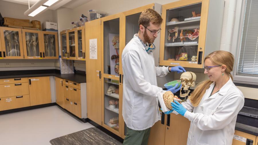
“Furthermore, it complements the Cadaver Lab, as students are now able to observe both microscopic (cell/tissue-level) anatomy and gross (organ-level) anatomy.”
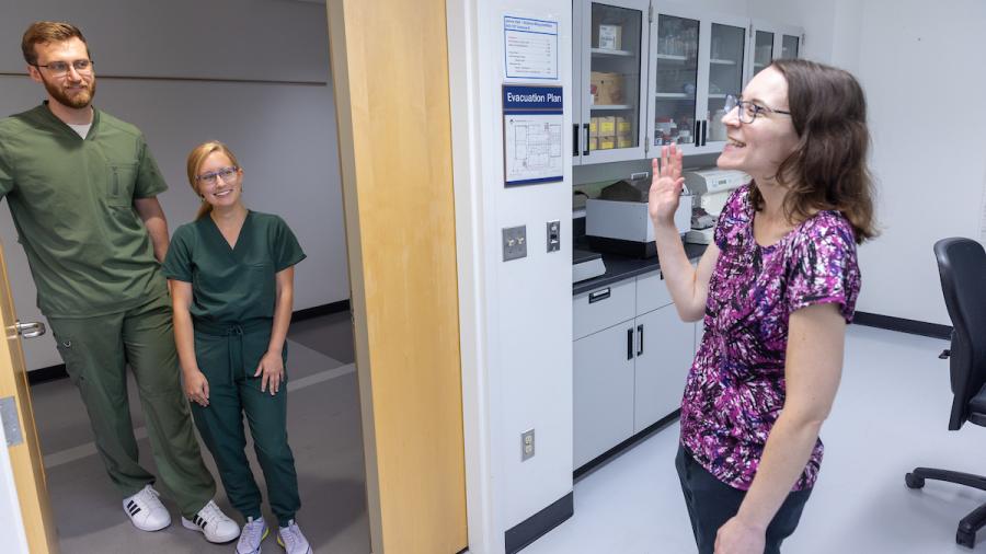
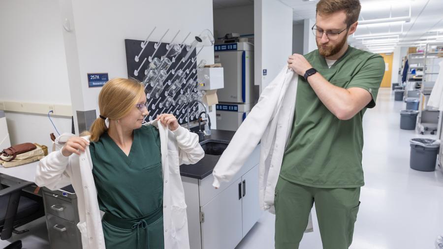
Last spring semester, Hoage worked with seniors Ethan Kalin and Natalie Mercill to establish the space within Jarvis Hall Science Wing’s larger biotechnology labs.
Histology is the study of the microscopic structures of biological tissues and is important for medical diagnosis, forensic investigation and scientific study.
While UW-Stout’s Histology Lab is not intended to diagnose patients, it will help students prepare for their future careers in biological and biomedical research and health care fields.
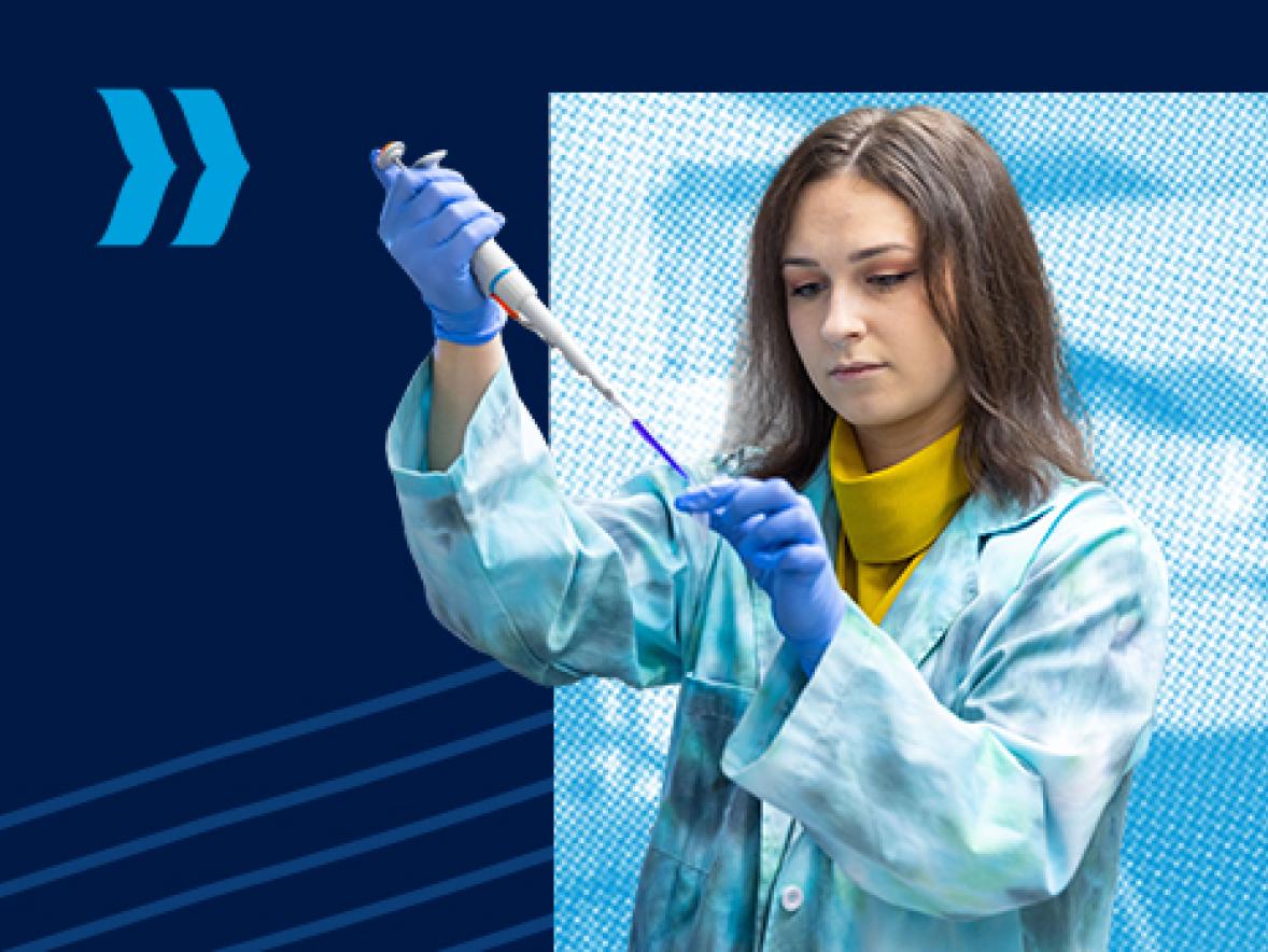
As they were establishing the Histology Lab, Kalin and Mercill learned how to operate the equipment, which includes a Leica EG1160 paraffin embedding center, a Leica RM2255 rotary microtome, a Fisher Scientific warm water bath and staining equipment.
They used such equipment only once before during a tour of Mayo Clinic’s histology department with the university’s Prehealth Society.
“The tour definitely got us interested in histology,” Mercill said.
In May, Kalin, of Maplewood, Minn., graduated from UW-Stout in applied science, with a concentration in biology and minors in cognitive neuroscience and chemistry. Mercill, of Woodbury, Minn., graduated in biology from UW-Eau Claire.
They now work at Mayo Clinic in Rochester, Minn. Kalin is a clinical lab technologist in a genomics lab, extracting DNA for cancer and hereditary disease diagnosis. Mercill does phlebotomy as a lab services technician and is starting the cardiovascular invasive specialist program at the Mayo School of Health Science, where she will learn the skills to assist with diagnostic and therapeutic invasive cardiology procedures.
“Getting to work in the Histology Lab made me much more confident in my ability to pick up new techniques and put them into practice. I believe this has helped me work through training I have undergone at my new position in Rochester. I hope to see the lab we created allow for other students to grow in the same ways I have and discover new passions that will help expand their horizons,” Kalin said.
Revisiting the lab before it opens for the semester, Kalin and Mercill practice what their peers will soon be experiencing in class.
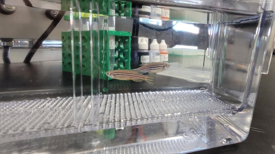
They extract the brain from a zebrafish and use the microwave method to dehydrate the tissue to prepare it for paraffin embedding. Zebrafish are a model organism for research, as the fish shares more than 80% of genes known to trigger diseases in humans, such as cancer types and heart disease.
“The brain of an adult zebrafish is about four millimeters in length, or the width of a sunflower seed. Even with that small of a tissue sample, you can create hundreds of microscope slides,” Mercill said.
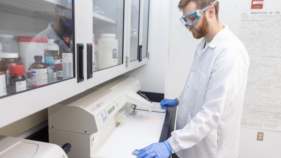
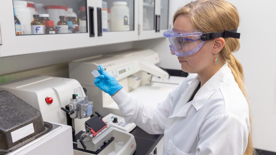
At the paraffin embedding center, Kalin places the silver mold under the paraffin wax dispenser and partially fills it. He uses a forceps to place the tissue into the molten wax.
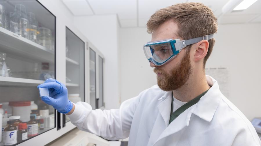
He places an embedding cassette, a blue plastic slotted tray about one-inch square, into the silver mold. Additional molten paraffin wax is added, and he places mold on a cooling platform to solidify the wax and form the tissue block.
Mercill readies the rotary microtome to slice the tissue block into sections around five microns thick. By comparison, an average human hair has a diameter of 100 microns.
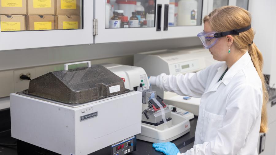
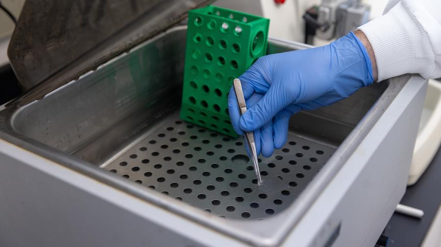
She clamps the tissue block into the microtome and installs a blade into the holder below. Turning the operating handle, she moves the block up and down to slice it, creating a length of perforated squares for microscope slides.
Mercill uses a pair of tweezers to lift a perforated section from the tray and hands it to Kalin, who lays it in the warm water bath, heated to 37 degrees Celsius, or 98.6 degrees Fahrenheit.
As it floats on top, the warm water flattens and smooths the section, and it is adhered to a slide and dried.
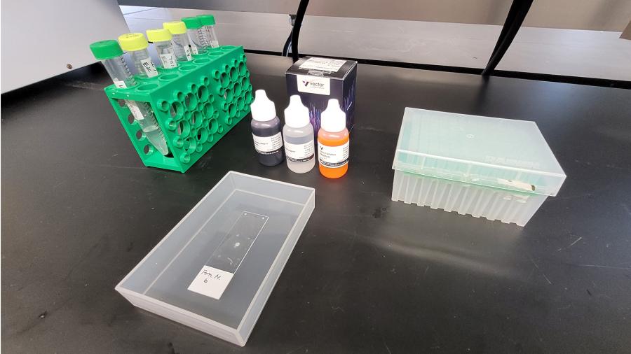
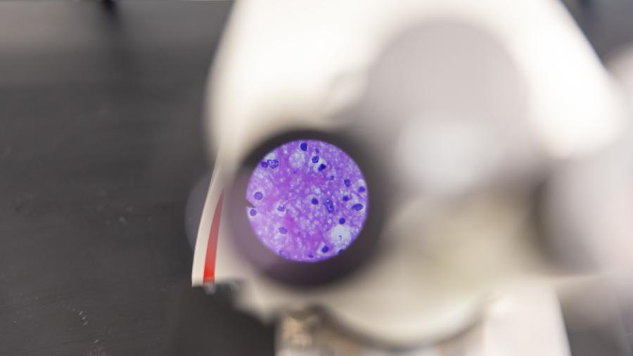
The sections are then stained to observe the tissue’s components. Specific stains are used to better visualize cells and structures of interest, Hoage explained, noting thioflavin-S highlights the amyloid beta, a protein that accumulates in the brains of patients with Alzheimer’s disease, while H&E – hematoxylin and eosin – is used to stain DNA purplish blue and the rest of the cell pink.
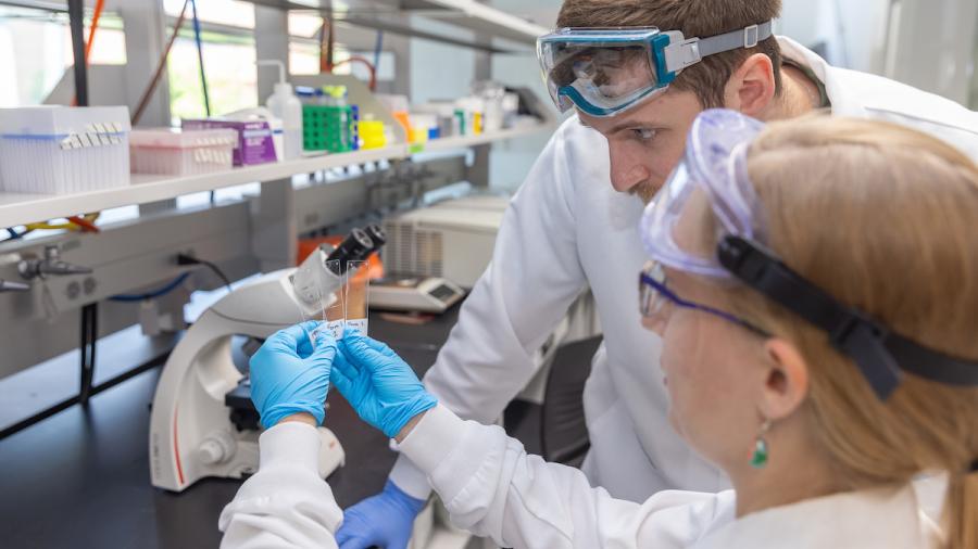
Under the microscope, Kalin and Mercill observe zebrafish tissue stained with H&E.
The initiative to establish the lab stemmed from an Honors College project in fall 2023 that involved Kalin and Mercill dissecting a brain from a cadaver with Alzheimer’s disease. They wanted to further their educational experience and see the microscopic pathology of Alzheimer’s disease, so their mentor Dr. Alex Hall connected them with Hoage.
With a Ph.D. in biomedical sciences from Mayo Clinic Graduate School and histology experience from her thesis research, Hoage was thrilled at the opportunity to help establish the Histology Lab.
“With Ethan and Natalie helping to set up the lab and now working at Mayo, it feels like they’re completing the cycle,” Hoage said.
The lab allowed them to continue their project at the microscopic level when they successfully sectioned and stained the cadaver brain to see amyloid beta plaques.
In another project, Kalin and Mercill compared the conventional method of tissue processing with the microwave method for processing zebrafish tissues.
“Their preliminary findings show both tissue processing methods are equivalent with respect to tissue integrity and staining quality,” Hoage said. “Their findings are important, as the microwave method takes less than one hour versus the conventional method, which takes about six hours. The shorter processing time will allow the microwave method to be integrated within a regular lab period.”
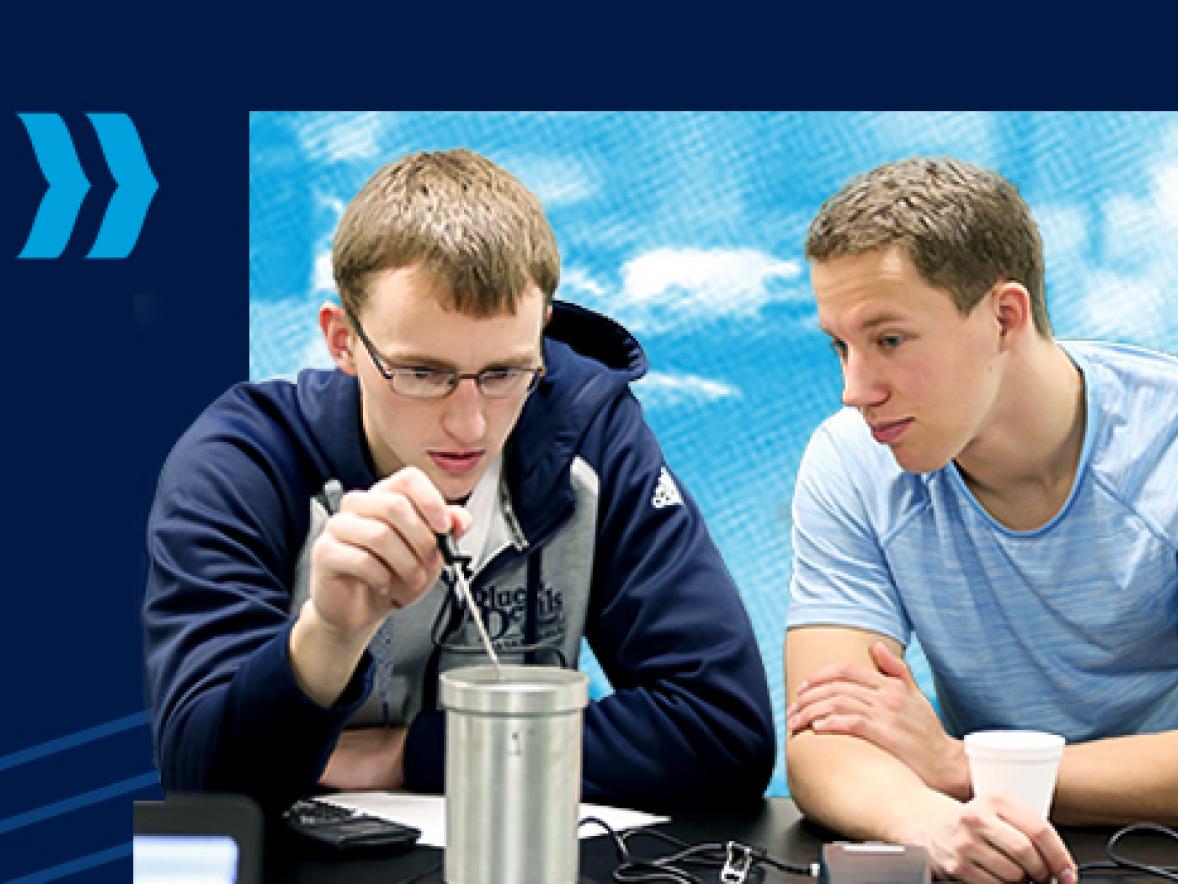
Kalin and Mercill were among nearly 300 undergraduate and graduate students who presented at UW-Stout’s Research Day in April. Kalin also presented his temporal analysis at Research in the Rotunda in March at the state Capitol.
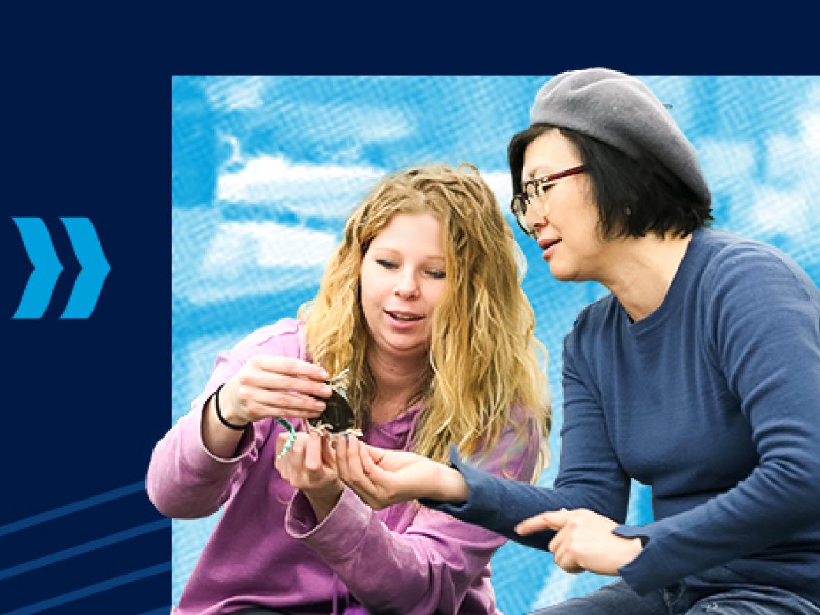
During their undergrad, Kalin and Mercill volunteered in the Cadaver Lab with Hall, giving tours to visiting high school students. Nearly 300 students from 15 high schools visited last semester to examine human organ systems on the 75-minute tour.
UW-Stout’s Pre-Health Pathways prepares students to enter medical school and other health care fields, including chiropractic, dentistry, pharmacy, medicine, occupational therapy, veterinary medicine and more.
Pre-health degrees include applied biochemistry and molecular biology; biology; health, wellness and fitness; rehabilitation services ; applied science, with an interdisciplinary science concentration; dietetics; environmental science with an environmental health concentration; human development and family studies; and psychology.
The biology, chemistry and physics majors have branched out of the applied science program, giving students the option to focus their degree on their science of choice.
The Office of Research and Sponsored Programs and the Stout University Foundation support student research and dissemination projects through internal and external grant programs.